
Healthcare with Confidence
Glioma and Glioblastoma Surgery in Israel: How a Neurosurgeon Chooses the Safest Treatment Approach
Below you will find clear information about glioma and glioblastoma surgery in Israel, including how neurosurgeons decide whether surgery or biopsy is safer, which advanced technologies may be used in Israel, and what treatment may follow.
You will also learn when to seek a second opinion, what documents and MRI scans to send, and how an Israeli medical team can help you plan every stage of care from abroad.
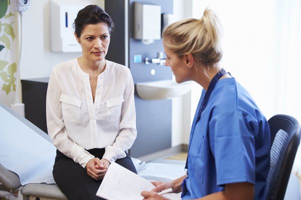
If you have been diagnosed with a glioma or glioblastoma, one of the most important questions is whether the tumor can be removed and how safely the surgery can be performed.
There is no simple answer. Gliomas often do not have clear borders and may be located close to areas of the brain responsible for speech, movement, memory, vision, or important neural pathways.
For this reason, modern glioma surgery is not simply about “removing the tumor.” It requires a careful balance between two equally important goals:
- removing as much of the tumor as safely possible;
- preserving your neurological functions, independence, and quality of life.
In Israel, surgery for glioma and glioblastoma is planned by a multidisciplinary medical team that may include a neurosurgeon, neuro-oncologist, neuroradiologist, neuropathologist, and radiation oncologist.
This team-based approach helps doctors understand in advance what surgery may achieve, which risks should be considered, and what additional treatment you may need after the operation.
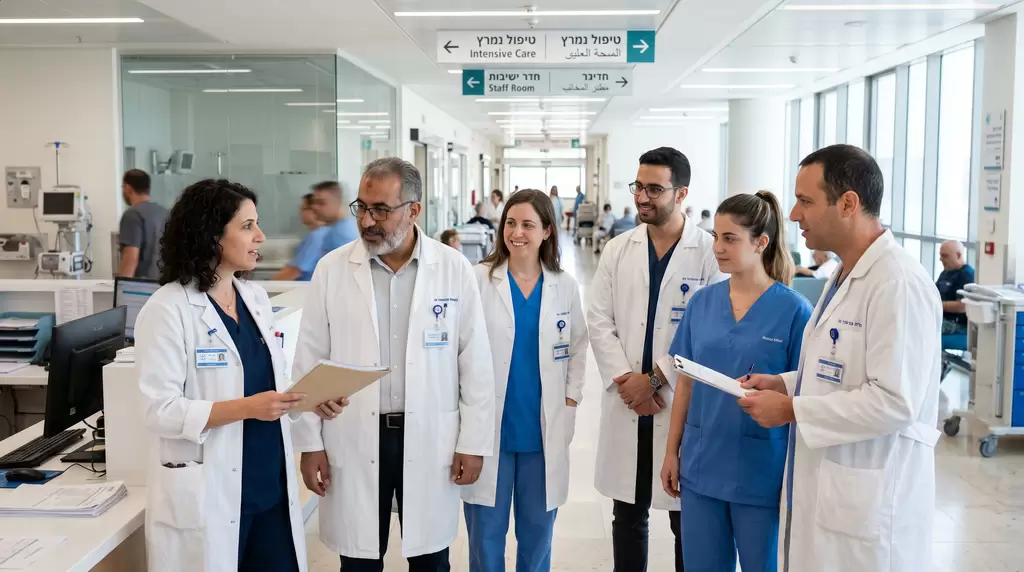
⇒ Best Israeli Neurosurgeons – Consultation
⇓ Below, you will find detailed information about some of the most experienced neurosurgeons in Israel
What Your Doctor Needs to Determine Before Surgery
Before discussing the date of surgery, your doctor needs to answer several important questions.
Where Is the Tumor Located?
One glioma may be located in a relatively accessible area of the brain. Another may be close to the speech center, motor cortex, visual pathways, or brainstem.
The tumor’s location affects the surgical approach, how much of it can be removed, and the risk of complications.
What Type of Glioma Is It?
It is not enough to define the tumor simply as a “glioma” or “glioblastoma.” Your medical team may also evaluate important molecular and genetic features, including:
- IDH-mutant or IDH-wildtype status;
- MGMT promoter methylation;
- ATRX;
- TP53;
- TERT;
- EGFR;
- CDKN2A/B;
- 1p/19q codeletion;
- other markers when necessary.
These results help doctors understand how the tumor may behave and which treatment may be recommended after surgery.
Do You Have Symptoms?
Seizures, weakness, speech difficulties, vision problems, headaches, drowsiness, or changes in behavior may influence how urgently surgery is needed.
Can the Tumor Be Removed Safely?
In some cases, the neurosurgeon can plan an extensive tumor resection. In others, removing only part of the tumor may be safer.
Sometimes, the first step is a biopsy rather than major brain surgery.
What Treatment Will You Need After Surgery?
For glioblastoma, surgery is almost always only the first stage of treatment. After the operation, your medical team will usually discuss radiotherapy, temozolomide chemotherapy, additional treatment options, and follow-up MRI scans.
A well-designed surgical plan therefore takes the next stage of treatment into account from the beginning.
Glioma and Glioblastoma: Why the Surgical Approach Is Different
Glioma is a broad group of brain tumors. Glioblastoma is one of the most aggressive forms of diffuse glioma in adults.
Surgery for Low-Grade Glioma
For a low-grade glioma, surgery may be performed to achieve long-term tumor control, reduce the risk of progression, manage seizures, and obtain an accurate diagnosis.
When it is safe, the neurosurgeon may try to remove as much of the tumor as possible.
Surgery for High-Grade Glioma
For a high-grade glioma, the goal is usually to remove the maximum safe amount of active tumor, obtain tissue for detailed analysis, and prepare you for further treatment.
Surgery for Glioblastoma
Glioblastoma surgery is usually performed to reduce the tumor burden, relieve pressure on the brain, confirm the diagnosis, and improve the conditions for radiotherapy and chemotherapy.
However, further treatment is still required even after a successful resection because microscopic tumor cells may remain in the surrounding brain tissue.
When Is Surgery Needed, and When Is a Biopsy Enough?
Major brain surgery is not always the best first step.
Surgery May Be Recommended If:
- the tumor can be removed safely;
- the tumor is putting pressure on the brain;
- there is significant brain swelling;
- neurological symptoms are getting worse;
- reducing the tumor size may relieve symptoms;
- the tumor is causing seizures;
- glioblastoma is suspected;
- enough tissue is needed for an accurate pathological and molecular diagnosis.
A Biopsy May Be Preferred If:
- the tumor is located deep within the brain;
- surgery carries a high risk of neurological complications;
- the tumor involves critical areas responsible for speech, movement, vision, or other essential functions;
- the main goal is to confirm the diagnosis before radiotherapy or drug treatment;
- your general condition does not allow major surgery;
- the diagnosis remains uncertain.
A biopsy should not be viewed as “less treatment.” In some situations, it is the safest and most appropriate way to obtain an accurate diagnosis and begin the right therapy without unnecessary risk.
What Does “Maximum Safe Resection” Mean?
Maximum safe resection is one of the main principles of modern glioma surgery.
It means that the neurosurgeon removes as much of the tumor as possible without creating an unjustified risk to your speech, movement, vision, memory, independence, or overall quality of life.
Sometimes, a postoperative MRI shows that all visible tumor has been removed. In other cases, a small part of the tumor is intentionally left behind because removing it could cause a serious neurological deficit.
This is not always a surgical failure. In some situations, leaving a small amount of tumor is the safest and most appropriate decision.
A successful operation is not defined by the MRI image alone. It is also measured by how you feel and function after surgery: whether you can speak, move, understand, walk, eat, communicate, remain independent, and continue with the next stage of treatment.
How Glioma Surgery Is Planned in Israel
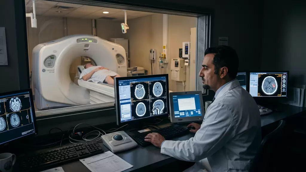
Glioma surgery does not begin in the operating room. It begins with a detailed analysis of your brain scans and neurological functions.
- MRI With Contrast. Your doctor evaluates the size of the tumor, contrast enhancement, swelling, necrosis, exact location, and its relationship to critical areas of the brain.
- Functional MRI. Functional MRI helps identify areas responsible for speech, movement, and other important functions. This is especially important when the tumor is located in the frontal, temporal, or parietal lobe, or close to the language areas of the brain.
- DTI Tractography. DTI tractography shows the major nerve pathways in the brain. For the neurosurgeon, it works like a map of important roads that must be protected during surgery.
- MR Perfusion and Additional MRI Sequences. MR perfusion and other advanced MRI techniques may help assess the activity of the tumor, especially when it is difficult to distinguish between tumor growth, swelling, and treatment-related changes.
- Review of Previous MRI Scans. If you have previous MRI scans, your doctor will compare them with the latest images. This is particularly important for gliomas because the speed and pattern of tumor growth may influence the treatment strategy and urgency of surgery.
Technologies That Help Israeli Neurosurgeons During Surgery
Modern glioma and glioblastoma surgery may involve several advanced technologies. These tools do not replace the experience of the neurosurgeon, but they help make the procedure more accurate and safer.
- Neuronavigation. Neuronavigation helps the surgeon use your MRI data as a three-dimensional map during the operation. It is especially valuable for deep brain tumors or tumors with poorly defined borders.
- 5-ALA Fluorescence-Guided Surgery. Before surgery, you may receive a medication called 5-ALA. Under a special light, certain tumor cells become fluorescent and easier to distinguish from healthy brain tissue. This technology may help the neurosurgeon identify active tumor tissue more clearly, particularly in high-grade gliomas and glioblastoma.
- Intraoperative MRI. Intraoperative MRI allows the surgical team to assess the result while the operation is still in progress. When visible tumor remains and can be removed safely, the neurosurgeon may continue the resection during the same procedure.
- Intraoperative Ultrasound. Intraoperative ultrasound may be used to monitor the location of the tumor and changes in the brain after the resection has begun. It can provide the surgeon with updated information during the operation.
- Cortical and Subcortical Brain Mapping. The neurosurgeon may stimulate specific areas of the brain and important neural pathways to identify where essential functions are located. This helps define the boundaries of safe tumor removal and protect areas responsible for speech, movement, and other neurological abilities.
- Intraoperative Neurophysiological Monitoring. During surgery, the medical team may continuously monitor the function of the nervous system. This helps reduce the risk of injury to the motor pathways and other critical brain structures.
- Awake Craniotomy. When the tumor is located close to areas responsible for speech or complex cognitive functions, an awake craniotomy may be recommended. During part of the operation, you may be asked to complete simple tasks, such as speaking, naming objects, counting, or moving your hand. This is not done to test your endurance. It allows the surgical team to monitor and protect your brain functions in real time. You should not feel pain during the procedure, and an experienced anesthesia team supports and monitors you throughout every stage of the operation.
What Happens During the First Days After Glioma Surgery?
After brain tumor surgery, it is not enough simply to “wake up.” Your medical team needs to assess how safely the procedure was completed, how your brain is functioning, and what the next stage of treatment should be.
Follow-Up MRI
A postoperative MRI is usually performed to determine how much of the tumor was removed and whether any visible tumor tissue remains.
The scan also helps doctors assess swelling, bleeding, and other early postoperative changes.
Neurological Assessment
Your medical team will evaluate your speech, movement, sensation, vision, memory, coordination, and level of consciousness.
Some temporary neurological changes may occur after surgery because of swelling or manipulation of brain tissue. Your doctors will monitor these changes closely and determine whether rehabilitation or additional treatment is needed.
Management of Brain Swelling
Brain swelling may remain or temporarily increase after surgery.
Your doctor may prescribe corticosteroids or other medications to control the swelling and gradually reduce the dose when it is safe to do so.
Prevention and Treatment of Seizures
If you experienced seizures before surgery, anticonvulsant treatment is especially important.
When there was no history of seizures, the decision to use preventive medication is made individually according to the tumor location, type of surgery, and your overall condition.
Pathology Results
The final diagnosis is based on examination of the tumor tissue removed during surgery.
A preliminary pathology result may be available earlier, while the complete molecular and genetic analysis usually takes longer.
Why Tumor Tissue Is So Important
In glioma and glioblastoma treatment, surgery is not performed only to remove the tumor. It also provides tissue for an accurate diagnosis.
Pathological and molecular testing may determine:
- the exact type of brain tumor;
- the tumor grade;
- IDH status;
- MGMT promoter methylation;
- 1p/19q codeletion;
- mutations and other molecular changes that may influence treatment;
- signs associated with more aggressive tumor behavior.
Without this information, treatment planning may be less precise.
For this reason, if your surgery was performed in another country, Israeli specialists may recommend a pathology review of the original slides and paraffin blocks before confirming the diagnosis or recommending further treatment.
Treatment After Surgery: What Is Usually Discussed?
After surgery for a glioma or glioblastoma, the next treatment plan depends on the final diagnosis, tumor grade, molecular profile, extent of resection, and your overall condition.
After Glioblastoma Surgery
Treatment usually includes radiotherapy combined with temozolomide, followed by additional cycles of temozolomide.
In selected cases, your medical team may also discuss Tumor Treating Fields, clinical trials, targeted options based on molecular findings, or other treatment approaches.
For IDH-Mutant Astrocytoma
The treatment strategy depends on several factors, including:
- the tumor grade;
- your age and general condition;
- how much of the tumor was removed;
- your symptoms;
- the tumor’s molecular features.
Possible approaches may include active surveillance, radiotherapy, chemotherapy, or combined treatment.
For Oligodendroglioma
When an IDH mutation and 1p/19q codeletion are confirmed, the treatment approach may differ from the plan used for astrocytoma.
Depending on the tumor grade, your symptoms, and the extent of surgery, treatment may include observation, radiotherapy, chemotherapy with the PCV regimen, or another individualized plan.
If the Tumor Returns
For recurrent glioma or glioblastoma, the medical team may discuss:
- repeat brain tumor surgery;
- repeat radiotherapy in selected cases;
- drug treatment;
- Tumor Treating Fields;
- participation in clinical trials;
- rehabilitation and supportive care.
The best option depends on the location and size of the recurrence, previous treatment, molecular findings, neurological condition, and the time since the original therapy.
Conservative Technologies and Less Invasive Treatment Decisions
Modern treatment does not always mean doing more. In some situations, the most advanced approach is to avoid unnecessary intervention and choose the safest option for you.
Active Surveillance
After surgery for certain low-grade gliomas, or when a small lesion remains stable, your doctor may recommend regular follow-up with MRI scans rather than immediate additional treatment.
Safe Partial Resection
When complete tumor removal could cause a serious neurological deficit, the neurosurgeon may remove only the part that can be treated safely.
The goal is to reduce the tumor burden while protecting your speech, movement, vision, memory, and independence.
Biopsy Instead of Major Surgery
When the tumor is located deep in the brain or within a critical functional area, a biopsy may be safer and more appropriate than extensive surgery.
The tissue obtained can still provide the diagnosis and molecular information needed to plan radiotherapy or drug treatment.
Symptom Management
Treatment of brain swelling, seizures, pain, sleep difficulties, anxiety, and other symptoms can significantly improve how you feel and help prepare you for the main treatment.
Rehabilitation
Physiotherapy, speech therapy, occupational therapy, and neuropsychological support may help you regain neurological functions, adapt to changes, and improve your quality of life after surgery.
Supportive Care
With an aggressive brain tumor, it is important to discuss not only how the tumor will be treated, but also how you will feel during therapy.
Supportive care may address nutrition, sleep, fatigue, medication side effects, infection risk, emotional well-being, and practical support for you and your family.
When Glioblastoma Is Located in a Complex Area of the Brain
Some glioblastomas are located close to critical brain regions that must be protected during surgery.
Near the Speech Areas
If the tumor is close to the areas responsible for speech and language, an awake craniotomy with speech mapping may be recommended.
This allows the surgical team to monitor your ability to speak, understand words, and name objects while the tumor is being removed.
Near the Motor Area
When the tumor is close to the areas controlling movement, the neurosurgeon may use intraoperative neurophysiological monitoring, brain mapping, and DTI tractography.
These technologies help identify and protect the pathways responsible for movement.
Near the Visual Pathways
The neurosurgeon will carefully assess the risk of vision loss or deterioration. In some cases, the extent of tumor removal may be limited to protect your eyesight.
In Deep Brain Structures
When the tumor is located deep within the brain, a biopsy or partial resection may be safer than extensive surgery.
The goal is to obtain an accurate diagnosis and reduce the tumor burden without creating an unacceptable risk of neurological damage.
In the Corpus Callosum or With Bilateral Spread
When glioblastoma involves the corpus callosum or spreads to both sides of the brain, surgery may be limited.
In these situations, the main focus of treatment may shift toward radiotherapy, chemotherapy, Tumor Treating Fields, clinical trials, or other individualized approaches.
An experienced neurosurgeon will not promise complete tumor removal in every situation. Instead, the surgeon should clearly explain where the boundary of safe resection lies and why a more limited procedure may sometimes be the right decision.
When Should You Get a Second Opinion Before Glioma or Glioblastoma Surgery?
A second neurosurgical opinion is especially important if:
- you have been advised to undergo urgent surgery but do not fully understand its purpose;
- the tumor is located close to critical areas of the brain;
- only a biopsy has been recommended and you want to know whether tumor removal may be possible;
- surgery has been proposed without a clear explanation of the risks and expected benefits;
- the diagnosis remains uncertain;
- doctors are considering whether the lesion is a glioma, brain metastasis, lymphoma, or an inflammatory process;
- you have already had surgery, but visible tumor remains;
- a repeat operation is being considered because the tumor has returned;
- you want to understand which advanced surgical technologies may be available in Israel.
A second opinion does not always change the treatment plan. Sometimes, it confirms that the proposed approach is appropriate.
However, confirmation from an experienced specialist can give you greater confidence, clarity, and peace of mind before making an important decision.
It is essential to send the original MRI images, not only the written radiology report. For a neurosurgeon, the scans themselves are often more informative than the report.
Why People Choose Glioma and Glioblastoma Surgery in Israel
Brain tumor surgery requires extensive neurosurgical experience, advanced technology, and a highly skilled multidisciplinary team. This is especially important when the tumor is located close to critical areas of the brain or when the diagnosis requires precise pathological and molecular evaluation.
In Israel, you can receive:
- a consultation with a neurosurgeon specializing in brain tumors;
- an expert review of your MRI scans;
- an assessment of whether maximum safe tumor removal is possible;
- a surgical plan using advanced technologies when appropriate;
- a neuro-oncology consultation;
- a review of pathology slides and paraffin blocks;
- molecular and genetic testing of the tumor;
- a plan for radiotherapy and chemotherapy after surgery;
- support throughout your arrival, hospitalization, surgery, and treatment in Israel.
For you, this means more than simply traveling abroad for an operation. You receive a clear and coordinated treatment plan explaining what should be done before surgery, which risks need to be considered, what treatment may be required afterward, and how your follow-up should continue.
How We Can Help You
If you have been diagnosed with a glioma or glioblastoma, we can help arrange a consultation with an Israeli neurosurgeon or neuro-oncologist.
You can begin with an online second opinion by sending your MRI scans, medical reports, pathology results, and other relevant documents.
We can help you:
- understand whether surgery may be possible;
- assess how urgently treatment is needed;
- send your MRI images to an Israeli brain tumor specialist;
- receive an expert opinion about the safest surgical approach;
- clarify which surgical technologies may be useful in your case;
- arrange a pathology review and molecular testing;
- prepare your visit to Israel if surgery is recommended;
- receive support during your consultation, hospitalization, surgery, and further treatment.
Consult an Israeli Specialist About Glioma or Glioblastoma Surgery
Contact us to receive an expert online consultation regarding glioma or glioblastoma surgery in Israel.
You will receive a clear explanation of your diagnosis, an assessment of whether the tumor can be removed safely, and a personalized plan of action—from an online second opinion to treatment and surgery in Israel.
 Professor Zvi Ram
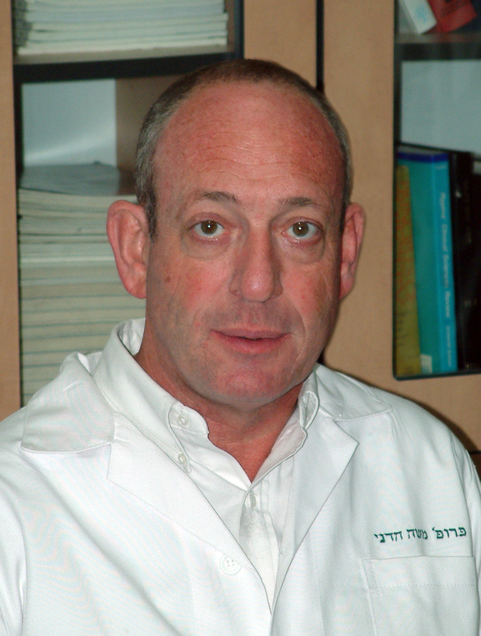 Professor Moshe Hadani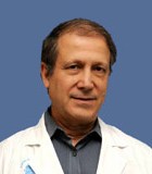
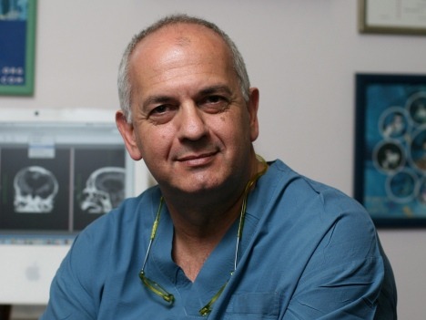 Professor Shlomo Constantini