
Healthcare with Confidence
Modern Methods of Meningioma treatment and Experienced Neurosurgeons
A diagnosis of meningioma can be alarming, especially if the tumor is located near the optic nerves, major blood vessels, the brainstem, or other critical structures.
However, meningiomas often grow slowly, and many are benign. Therefore, surgery is not required in every case.
In Israel, treatment decisions are made on an individual basis. The neurosurgeon evaluates the tumor’s location and size, its growth rate, your symptoms, your age, your general health, and the potential risks of intervention. Our doctors’ primary goal is to find the safest option while preserving your quality of life.
The guiding principle of Israeli neurosurgery is not to remove the tumor at any cost, but to achieve the best possible outcome while minimizing risk to vital functions.
What Is a Meningioma?
A meningioma is a tumor that develops from the membranes (meninges) covering the brain or spinal cord. It does not originate from the brain tissue itself, but as it enlarges, it can compress the brain, cranial nerves, and blood vessels.
Small meningiomas are sometimes discovered incidentally during an MRI scan and cause no symptoms. In such cases, a doctor might recommend monitoring rather than immediate surgery.
Is Surgery Always Necessary?
Our doctors consider surgery if the tumor:
- causes neurological symptoms;
- shows growth on follow-up MRI scans;
- compresses the brain or causes significant swelling;
- impairs vision, hearing, movement, or speech;
- is large in size;
- is located in a way that further growth could make treatment more difficult;
- appears aggressive based on diagnostic findings.
If a meningioma is small, slow-growing, and asymptomatic, an Israeli neurosurgeon may suggest active monitoring involving regular MRI scans.
International guidelines also recognize monitoring as a valid approach for asymptomatic tumors, whereas surgical removal is typically the primary treatment for growing or symptomatic meningiomas.
How the decision to operate is made in Israel
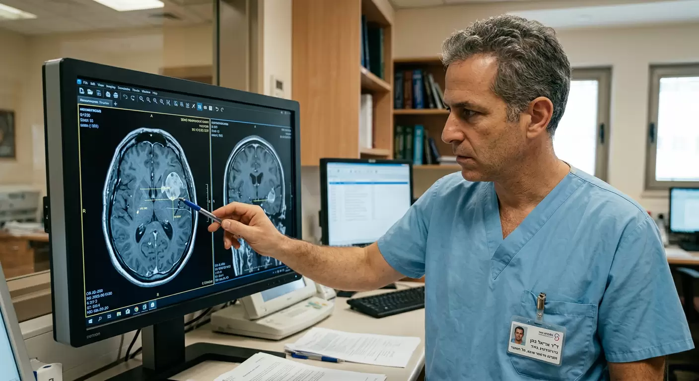
Before surgery, an Israeli neurosurgeon carefully examines not only the size of the tumor but also its relationship with surrounding structures.
Of particular importance are:
- contact with major arteries and venous sinuses;
- proximity to the optic nerves and chiasm;
- location near the brainstem;
- involvement of cranial nerves;
- degree of brain swelling (edema);
- history of prior surgeries or radiation therapy;
- likelihood of complete and safe removal.
Sometimes, attempting to remove the entire tumor can result in damage to a nerve or blood vessel. In such cases, the surgeon may leave a small fragment of the meningioma and recommend monitoring or precise radiation therapy.
Types of meningioma surgery performed in Israel
Microsurgical removal
This is the primary treatment method for most surgically accessible meningiomas. Through a carefully planned approach, the surgeon separates the tumor from the brain, nerves, and blood vessels using a surgical microscope and micro-instruments.
The size of the incision and the bone opening depends on the tumor’s location. A large meningioma does not always require a large incision; the surgeon selects the most direct and safe route.
Minimally invasive surgery
For certain tumors, a small surgical opening—known as “keyhole surgery”—may be used. This allows access to the meningioma through a limited opening in the skull.
This approach is not suitable for everyone. Its feasibility depends on the tumor’s size, growth pattern, and location. A minimal incision must not compromise the safety of the procedure.
Endoscopic transnasal surgery
Some skull base meningiomas can be accessed through the nasal passages. During an endoscopic endonasal procedure, a neurosurgeon and an ENT surgeon work together without making a traditional incision on the scalp.
This method may be considered for certain skull base tumors—for example, those located near the sella turcica and the optic nerves.
The choice between endoscopic and open surgery depends on the specific anatomy of the meningioma. Modern surgical approaches also include standard craniotomy, minimally invasive approaches, and endoscopic endonasal surgery for select skull base tumors.
Surgery for Skull Base Meningioma
Skull base tumors are among the most complex to treat because they can encase blood vessels and cranial nerves responsible for vision, hearing, eye movement, swallowing, and facial expressions.
In such cases, the neurosurgeon’s specific expertise in skull base surgery is crucial. The procedure may be performed in collaboration with an ENT surgeon, a vascular neurosurgeon, an ophthalmologist, or a plastic surgeon.
Modern technologies used during surgery
Leading neurosurgical centers in Israel may employ the following technologies:
- Neuronavigation. This functions like a GPS for the neurosurgeon. The system aligns the position of surgical instruments with your MRI scans, helping to precisely define the tumor’s boundaries and select a safe approach to it.
- Intraoperative neurophysiological monitoring. During surgery, specialists monitor the function of nerve pathways and cranial nerves. This is particularly important if the meningioma is located near areas responsible for movement, hearing, vision, or facial function. If signal changes occur, the surgeon can adjust their actions to reduce the risk of neurological damage.
- Surgical microscope and endoscope. Modern optics provide a better view of small blood vessels, nerves, and tumor areas hidden behind anatomical structures. In some surgeries, the microscope and endoscope are used in combination.
- Intraoperative MRI. In certain centers and clinical situations, an MRI scan can be performed directly during surgery. This helps assess the extent of the tumor removal and clarify the location of critical structures. For example, the Department of Neurosurgery at Sheba Medical Center utilizes microsurgery, neuronavigation, minimally invasive neuroendoscopy, and intraoperative MRI in the treatment of benign brain tumors, including meningiomas.
- Preoperative embolization. If a large meningioma has a rich blood supply, embolization may be considered prior to surgery. Using a vascular catheter, the doctor blocks some of the blood vessels feeding the tumor to minimize potential blood loss during removal. This procedure is not suitable for everyone and is prescribed only after an assessment of angiography and vascular anatomy.
Can a meningioma be completely removed?
The possibility of complete removal depends on the tumor’s location. If the tumor separates easily from the brain, blood vessels, and nerves, the surgeon may remove it entirely, along with the section of the meninges (membrane) from which it originates.
If the meningioma invades a major venous sinus, encases an artery, or is tightly adherent to a cranial nerve, partial removal is sometimes the safer option. The remaining tissue can be monitored or treated with stereotactic radiosurgery.
Before surgery, it is important to ask the neurosurgeon not only about the feasibility of complete removal but also about the functional risks associated with such an attempt.
What happens to the tumor after surgery
The removed tissue is sent for histological examination. A pathologist determines the meningioma type and its grade of malignancy according to World Health Organization (WHO) classification.
Additional tests may include:
- immunohistochemical analysis;
- assessment of Ki-67 proliferative activity;
- molecular and genetic testing;
- analysis of changes affecting the risk of recurrence.
The pathology results help determine whether surgery alone is sufficient or if additional treatment is required.
When radiation therapy is needed after surgery
Radiation treatment may be considered if:
- the tumor was not completely removed;
- the meningioma has recurred;
- a higher-grade meningioma is identified;
- repeat surgery carries a high risk;
- residual tumor tissue is located near critical nerves or blood vessels.
Depending on the tumor’s size and location, stereotactic radiosurgery or fractionated radiation therapy may be used.
Radiosurgery is not a conventional operation; no incision is made. Precise beams of radiation are directed at the tumor to halt its further growth.
Recovery after meningioma removal
After surgery, you will remain under the observation of the neurosurgical team for a period of time. In the initial days, pain medication, treatments for brain swelling, and anti-seizure therapy may be prescribed.
The length of the hospital stay depends on the extent of the surgery and your condition. Recovery is generally faster after the removal of a small, accessible meningioma than after complex skull base surgery.
Temporary after-effects may include:
- fatigue;
- headache;
- swelling at the incision site;
- dizziness;
- reduced concentration;
- temporary worsening of neurological symptoms.
If necessary, a physical therapist, speech therapist, balance specialist, or occupational therapist will be involved in your recovery.
Before you return home, the doctor will explain when you can fly, work, drive, exercise, and resume taking previously prescribed medications.
Potential surgical risks
Risks depend primarily on the location of the meningioma, not just its size. This does not mean that complications will necessarily occur.
Our neurosurgeon will explain your specific individual risks and the expected benefits of the surgery during the consultation, ensuring you are fully informed and able to make the best decision.
Documents required for a consultation
To obtain a preliminary opinion from an Israeli neurosurgeon, you typically need to provide:
- Brain MRI with contrast (in DICOM format);
- MRI report;
- Previous scans to assess changes over time;
- Skull CT scan (if performed);
- Medical summary describing symptoms;
- List of current medications;
- Details of previous surgeries and pathology reports (if treatment has already been administered);
- Information on other medical conditions and past procedures.
The actual MRI images are particularly important to the neurosurgeon, not just the written report.
Questions to ask the neurosurgeon
During the consultation, it is important to clarify the following:
- Is surgery required now, or can the meningioma be monitored?
- What happens if treatment is delayed?
- Can the tumor be completely removed?
- Which nerves and blood vessels are located nearby?
- What surgical approach do you recommend?
- Is a minimally invasive or endoscopic procedure possible?
- Will neurophysiological monitoring be required?
- Which functions might be temporarily or permanently altered?
- Might radiosurgery or radiation therapy be necessary?
- How long will I need to stay in Israel?
- Why choose Israel for meningioma surgery?
Meningioma surgery requires not only state-of-the-art equipment but also an experienced team, especially if the tumor is located at the skull base or near vital structures.
In Israel, you can access:
- Consultation with a neurosurgeon specializing in meningioma and brain tumors;
- Review of MRI scans and previous medical records;
- Personalized choice between monitoring, surgery, and radiosurgery;
- Microsurgical or endoscopic treatment;
- Neuronavigation and nervous system function monitoring;
- Analysis of tumor tissue;
- Collaborative decision-making involving a neurosurgeon, radiation oncologist, and neuro-oncologist;
- A follow-up plan for after you return home.
Neurosurgery departments at major Israeli medical centers treat brain tumors and accept both Israeli residents and patients traveling from abroad.
Neurosurgical consultation in Israel
If you have been diagnosed with a meningioma, you can obtain a professional opinion from an Israeli neurosurgeon before making a final decision.
The doctor will review your MRI scans and medical records, then assess the need for surgery, the extent of potential tumor removal, associated risks, and alternative treatment options. Following the consultation, you will receive recommendations to help you decide whether to travel to Israel for further diagnostics or surgical treatment.
Contact us and submit your MRI scans in DICOM format. We will arrange a consultation with a suitable Israeli neurosurgeon and, should surgery be recommended, assist in planning the necessary examinations, hospitalization, and treatment in Israel.
 Professor Zvi Ram
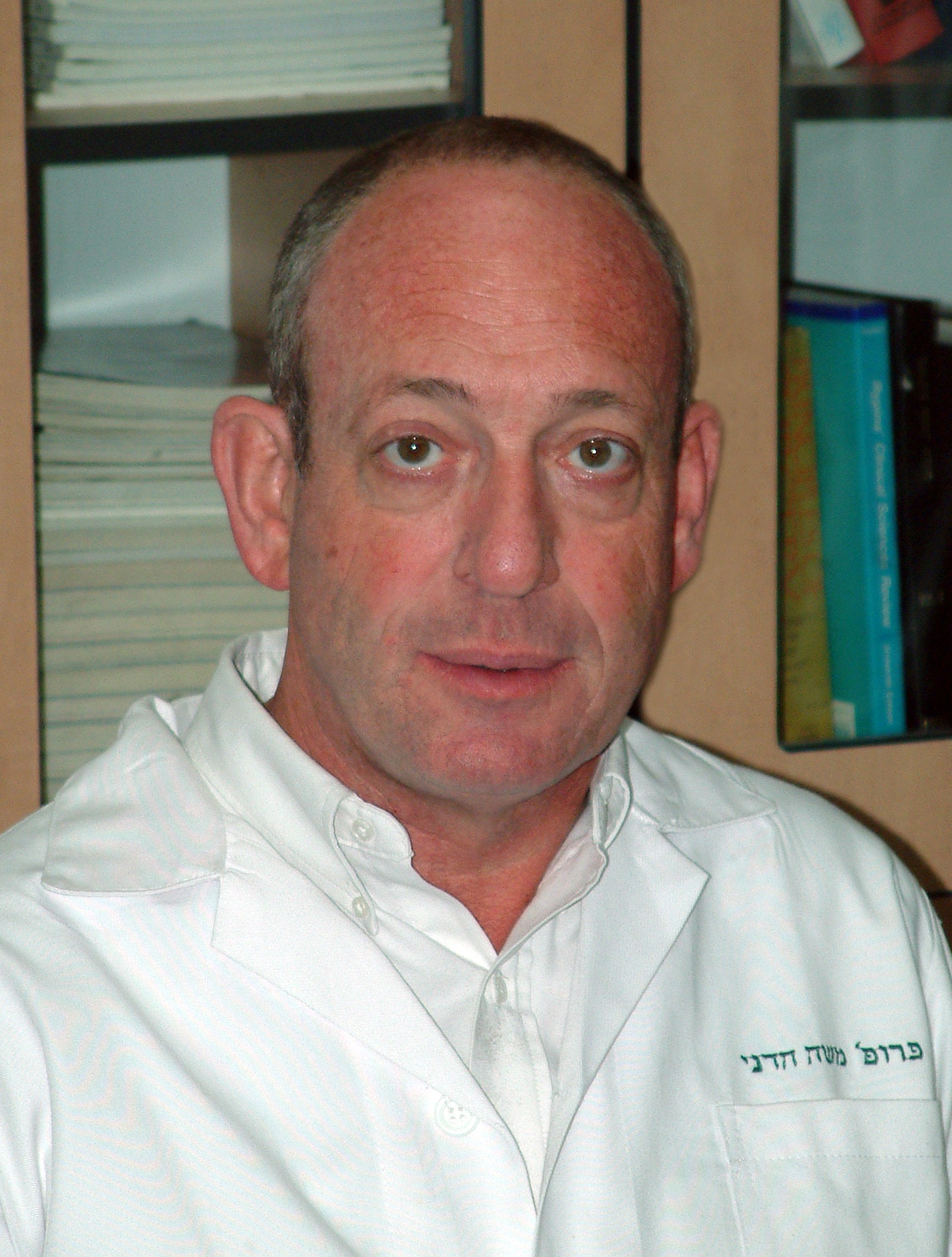 Professor Moshe Hadani
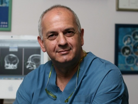 Professor Shlomo Constantini