Healthcare with Confidence
Kidney stones are crystalline solid concretion or aggregation formed in kidney from dietary minerals in the urine. Kidney stones typically leave the body with the flow of urine, and many stones are formed and leave without causing symptoms.
If stones enlarged to sufficient size ( at least 3 mm), they can cause obstruction (narrowing) of the ureter. Ureteral obstruction causes azotemia and hydronephrosis (distension and expansion of the renal pelvis and calyces) and also spasm of the ureter. This leads to pain, most commonly felt in the area between the ribs and hips), in the lower abdomen and groin (renal colic). Renal colic may also be associated with nausea, vomiting, fever, blood in the urine, pus and painful urination. Renal colic is usually wavy character and lasts 20 to 60 minutes, starting from the lower back and often irradiate to genitals.
Risk Factors
Dehydration from low fluid intake is a major factor of stone formation. High intake of animal protein, sodium, refined sugars, fructose, grapefruit and apple juice increases the risk of kidney stones. Kidney stones can cause distal renal tubular acidosis, Dent’s disease, hyperparathyroidism, primary hyperoxaluria. Kidney stones are more common in people with Crohn’s disease, which is associated with hyperoxaluria and magnesium absorption.
Calcium is one of the components of the most common type of kidney stones in man. Kidney stones develop and grow more in people who do not consume enough magnesium in the body. Magnesium inhibits the formation of stones. Evidence linking vitamin supplements C according to the studies are inconclusive. There is no conclusive evidence of cause – effect relationship between the consumption of alcoholic beverages and kidney stones. Nevertheless, there is an assumption that frequent alcohol drinking can lead to dehydration, which can in turn lead to the development of kidney stones.
When urine becomes supersaturated (solvent urine contains more solubles than can dissolve), small crystals are formed in kidney. The most common of them are formed of calcium oxalate, and they are usually the size of 4-5 mm.
Diagnostics
The diagnosis of kidney stones is based on history, physical examination, urinalysis, and radiographic studies, ultrasound examination and blood tests.
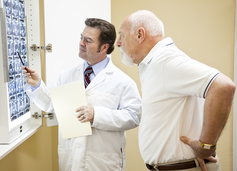
Radiographic examination of kidney stones carried by computed tomography (CT) with 5 millimeter slices. With this diagnosis all the stones can be revealed. The procedure can be executed with injection of a contrast agent or without it. Uroliths present in the kidneys, bladder or ureters may be better defined with use of a contrast agent.
Ultrasonography provides detailed information about hydronephrosis, it is assumed that the stone is blocking the flow of urine. Radiolucent stones that do not appear on CT scans can be displayed in ultrasound imaging. Ultrasound is useful for detecting stones in situations where x-rays or CT scans are not recommended, such as children and pregnant women. Despite these advantages, renal ultrasonography is not currently considered as a replacement for non-contrast helical computed tomography in the initial diagnostic evaluation of urolithiasis. The main reason for this is that, compared with CT, renal ultrasonography more often fails to detect small stones (especially ureteral stones) as well as other serious violations that could be causing the symptoms.
Laboratory studies usually include:
![]() microscopic examination of the urine, which may show red blood cells, bacteria, leukocytes, urinary casts and crystals
microscopic examination of the urine, which may show red blood cells, bacteria, leukocytes, urinary casts and crystals
![]() urine culture to detect any contamination of organisms present in the urinary tract as well as to determine the sensitivity of these organisms to specific antibiotics
urine culture to detect any contamination of organisms present in the urinary tract as well as to determine the sensitivity of these organisms to specific antibiotics
![]() common blood test to determine neutrophilia (increased neutrophil granulocytes), suggesting the presence of a bacterial infection
common blood test to determine neutrophilia (increased neutrophil granulocytes), suggesting the presence of a bacterial infection
![]() kidney function tests to determine whether the elevated levels of calcium in the blood (hypercalcemia)
kidney function tests to determine whether the elevated levels of calcium in the blood (hypercalcemia)
![]() 24 hour urine collection to measure total daily urinary volume, the content of magnesium, sodium, uric acid, calcium, citrate, oxalate and phosphate
24 hour urine collection to measure total daily urinary volume, the content of magnesium, sodium, uric acid, calcium, citrate, oxalate and phosphate
Treatment of Kidney Stones
Stone size affects the rate of stone spontaneous leaving the body. For example, up to 98 % of small rocks (at least 5 mm in diameter) can pass spontaneously through the urine within four weeks after the onset of symptoms, but for larger stones (5 to 10 mm) the rate of spontaneous passage decreases to less than 55 %. Initial stone location also influences the likelihood of spontaneous stone passage.
Analgesia therapy. For example, intravenous administration of opioids. Orally administered drugs are often effective for less severe discomfort and pains.
Medication removal of kidney stones shown to accelerate the spontaneous stone passage of the ureter. These procedures are also used in addition to the lithotripsy.
Extracorporeal shock wave lithotripsy (ESWL) is a noninvasive method of kidney stones removal, which is used when the stone is in renal pelvis. Lithotripter is a device that emits pulses of ultrasound energy focused high intensity, causing fragmentation of the stone for about 40-60 minutes. This method is used in the treatment of uncomplicated stones arranged in the upper part of the kidney and ureter with the proviso that the size of the stones smaller than 20 mm and normal kidney anatomy. Lithotripsy can not break a stone exceeding 10 mm in one session. Two or three treatments may be needed. Lithotripsy method can be effectively treated in from 85 % simple kidney stones. The effectiveness of this method affect a number of factors, including the chemical composition of the stone, abnormal renal anatomy and the specific location of the stone in the kidney, hydronephrosis, body mass index as well as the distance from the stone surface from the skin.
Surgical treatment
As already mentioned, most of the stones up to 5 mm pass spontaneously Surgery may be needed in those cases where there is only one working kidney, bilateral obstacle for stones, urinary tract infection.
Ureteroscopic surgery
Ureteroscopy is becoming an increasingly popular method of treatment, because this is the most gentle and effective. One method of ureteroscopy involves placing a ureteral stent (a small tube extending from the bladder to the ureter and kidney) to provide immediate relief. Stent may be useful to preserve kidneys from acute renal failure due to increased hydrostatic pressure, swelling and infection (pyelonephritis and pyonephrosis) caused by stone.
Holmium laser lithotripsy is a procedure that carries less risk than any other procedures. During this procedure performed under general anesthesia a catheter is inserted into the ureter, which is fed through a laser, fragmenting stones in the bladder, ureters and kidneys. Hospitalization for about 2 days.
We select the narrow section doctor who will determine the individual treatment in your case that makes treatment the most effective.


